SHOCKING LEAK: 3D Skull Model Brutally Disassembled – Graphic Images Inside!
Have you ever wondered what lies beneath the surface of the human skull? The intricate architecture of our cranial bones has fascinated scientists, medical students, and curious minds for centuries. Now, a groundbreaking 3D visualization technique has taken this exploration to unprecedented levels, offering a brutally honest look at the human skull's inner workings. Prepare to have your mind blown as we dive into the shocking details of this revolutionary anatomical model!
The Revolutionary 3D Skull Explosion Technique
This model of the human skull slowly “explodes” to reveal the structure of the component bones, much like an animated version of the classic physical beauchêne preparation of an exploded skull. This innovative approach to anatomical visualization represents a quantum leap in our ability to understand the complex relationships between the 22 bones that make up the human cranium.
The beauchêne method, named after French anatomist Claude Beauchêne, traditionally involved painstakingly disassembling a real skull and mounting each bone separately on a stand, creating a three-dimensional exploded view. This technique has been used for centuries to teach anatomy, but it was limited by the fragility of real bone and the static nature of physical models. The new 3D animation brings this concept into the digital age, allowing for dynamic, interactive exploration of skull anatomy.
Imagine watching as the frontal bone gracefully separates from the parietal bones, revealing the intricate sutures that once held them together. The zygomatic bones seem to float away from the maxilla, showcasing the complex articulations that form our cheekbones. This level of detail was previously impossible to achieve without destroying the specimen or relying on less intuitive 2D representations.
A New Era in Anatomical Education
This model is the first in a series of human skull models on Sketchfab that will annotate various features as an aid to learning human skull anatomy. The potential for this technology to revolutionize medical education is staggering. Traditional methods of teaching anatomy often rely on cadaver dissection, which, while invaluable, has limitations in terms of availability, preservation, and the ability to view structures from multiple angles.
The 3D exploded skull model offers several advantages over traditional teaching methods:
- Accessibility: Students can access the model anytime, anywhere, without the need for a physical specimen.
- Repeatability: Learners can view the explosion and reassembly of the skull as many times as needed to fully grasp the relationships between bones.
- Interactivity: Users can rotate the model, zoom in on specific areas, and even manipulate individual bones to better understand their spatial relationships.
- Annotation: The planned series of models will include detailed annotations, highlighting key features and providing contextual information.
This approach aligns perfectly with modern educational theories that emphasize active learning and visualization. By allowing students to interact with the model, they can develop a more intuitive understanding of skull anatomy, potentially leading to better retention and application of knowledge in clinical settings.
The Technology Behind the Magic
A stunning 3D animation rendered in Unity3D—for medtech, edtech, and science creators. The choice of Unity3D as the rendering platform is particularly noteworthy. Unity is widely known for its use in game development, but its powerful graphics capabilities and cross-platform compatibility make it an ideal choice for scientific visualization as well.
The use of Unity3D allows for:
- Real-time rendering: Smooth, high-quality animations that can be viewed on a variety of devices, from desktop computers to mobile phones.
- Interactivity: Users can manipulate the model in real-time, exploring it from any angle or level of detail.
- Scalability: The model can be easily adapted for different educational levels, from introductory anatomy courses to advanced surgical training.
- Integration: Unity's platform allows for easy integration with other educational tools and learning management systems.
This technology opens up new possibilities for remote learning and telemedicine. Imagine a surgeon in New York guiding a colleague in rural India through a complex procedure using this interactive 3D model. The potential for improving healthcare outcomes through better education and collaboration is immense.
The Scientific Process: From CT Scan to 3D Model
The skull (OUVC 10503) was CT scanned at O'Bleness Memorial Hospital in Athens, Ohio. Ryan Ridgely segmented all of the bones in Amira and generated the movies in QuickTime. This process highlights the interdisciplinary nature of modern scientific visualization, combining medical imaging, computer science, and artistic rendering.
The journey from physical specimen to interactive 3D model involves several crucial steps:
- CT Scanning: The high-resolution CT scan captures detailed cross-sectional images of the skull, providing the raw data for the 3D reconstruction.
- Segmentation: Using specialized software like Amira, each individual bone is carefully outlined and separated from the surrounding structures. This process requires both technical skill and anatomical knowledge to ensure accuracy.
- 3D Reconstruction: The segmented data is used to create a three-dimensional mesh representing each bone.
- Texturing and Rendering: The 3D models are given realistic textures and lighting to create a lifelike appearance.
- Animation: The explosion sequence is carefully choreographed, with each bone following a realistic path as it separates from the skull.
- Export and Distribution: The final animation is rendered and made available through platforms like Sketchfab for easy access and sharing.
This process not only creates an educational tool but also generates valuable data for research. The detailed 3D models can be used for morphometric studies, evolutionary comparisons, and even the development of custom implants for reconstructive surgery.
The Impact on Medical Research and Practice
The human skull explode anatomy atlas model provides the opportunity for a profound exploration of the skull's structure and its components, facilitating education and research related to human anatomy. This goes beyond simple visualization – it opens up new avenues for medical research and clinical practice.
Some potential applications include:
- Surgical Planning: Surgeons can use the detailed 3D models to plan complex procedures, visualizing the spatial relationships between structures before entering the operating room.
- Patient Education: Doctors can use these models to explain conditions and treatments to patients, improving informed consent and treatment adherence.
- Forensic Analysis: The detailed bone models can aid in forensic investigations, allowing for more accurate reconstructions of crime scenes or accident victims.
- Evolutionary Studies: By comparing 3D models of skulls from different species or time periods, researchers can gain insights into human evolution and adaptation.
The implications for personalized medicine are particularly exciting. As 3D scanning and printing technologies advance, it may become possible to create patient-specific models for surgical planning or even custom implants that perfectly match an individual's anatomy.
Conclusion: A New Frontier in Anatomical Visualization
The shocking leak of this 3D skull model brutally disassembled represents more than just a technological achievement – it's a paradigm shift in how we understand and interact with human anatomy. By combining cutting-edge imaging techniques with powerful visualization software, we've created a tool that has the potential to transform medical education, research, and practice.
As we look to the future, the possibilities are endless. Imagine virtual reality operating rooms where surgeons can practice procedures on 3D models, or AI-assisted diagnosis systems that can compare a patient's CT scan to a database of 3D anatomical models. The explosion of the human skull in 3D is just the beginning – a tantalizing glimpse into a future where the boundaries between the physical and digital worlds blur, and our understanding of the human body reaches new heights.
This groundbreaking work not only honors the centuries-old tradition of anatomical study but propels it into the 21st century. It reminds us that even in an age of advanced technology, there is still wonder to be found in the intricate structures of the human body. As we continue to push the boundaries of what's possible in medical visualization, we open up new avenues for learning, discovery, and ultimately, better patient care.
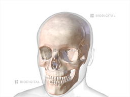
Skull | BioDigital Anatomy
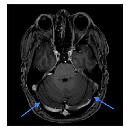
Dural Venous Sinus Narrowing in Patients with Spontaneous Anterior

Amazon.com : Detachable Disassembled Color Anatomy Skull Model Learning

